Fig. 6.
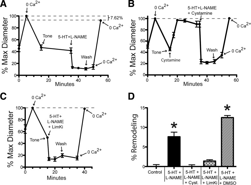
Exposure of isolated arterioles to 5-HT + l-NAME induces inward remodeling. After development of spontaneous myogenic tone, cannulated and pressurized arterioles were exposed to a 5-min incubation in 10−4 M adenosine + 0 Ca2+ + 2 mM EGTA (to establish maximum passive diameter), washed with fresh buffer for 20 min (to re-establish tone), exposed to vasoconstrictor agents for 10 min, washed with fresh buffer for 5 min, and exposed to 10−4 M adenosine + 0 Ca2+ + 2 mM EGTA (to re-assess maximum passive diameter). In A–C, the luminal diameter of arterioles is expressed as percentage of the initial maximal diameter obtained. A: diameter of arterioles exposed to 10−4 M 5-HT + 10−4 M l-NAME as vasoconstrictor agents. B: diameter of arterioles exposed to 10−4 M 5-HT + 10−4 M l-NAME + 10−3 M cystamine, including a 20-min incubation in 10−3 M cystamine before application of vasoconstrictor agents. C: diameter of arterioles exposed 10−4 M 5-HT + 10−4 M l-NAME + 10−6 M LimKi as vasoconstrictor agents. D: percent remodeling expressed as percent change in the maximal passive diameter measured after vs. that measured before exposure to vasoconstrictor agents. Data are means ± SE; n = 3 for 5-HT + l-NAME + DMSO, and for all other treatments n = 6. *P ≤ 0.05 vs. Control.

