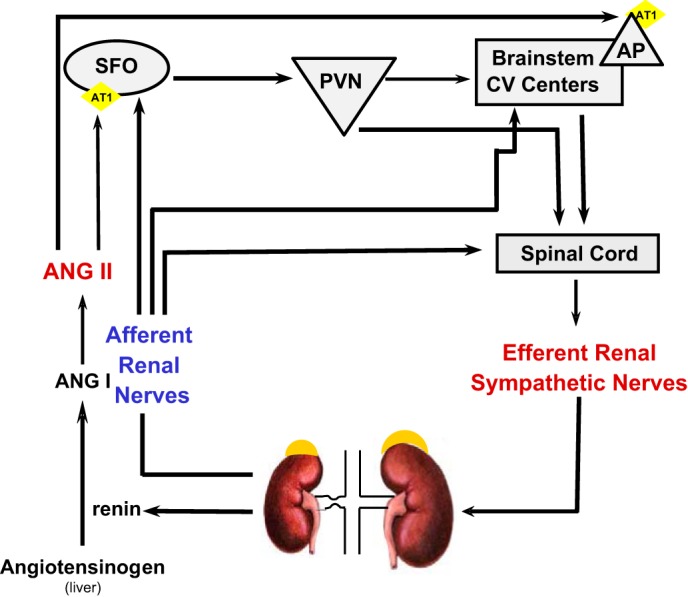
Fig. 10.

Diagram of potential mechanisms involved in increased ANG II and efferent RSNA to the contralateral kidney in 2K-1C hypertension. Hypoperfusion of the stenotic kidney results in increased secretion of renin, thereby initiating a cascade leading to elevated plasma ANG II. Plasma ANG II can act to increase vascular resistance directly (not shown) but can also act on brain nuclei that lie outside the blood-brain barrier and possess AT1 receptors such as the subfornical organ (SFO) or area postrema (AP). Projections from the AP or the SFO via paraventricular nucleus (PVN) can then send signals either via the brainstem cardiovascular regulatory centers or directly to the spinal cord to enhance efferent RSNA. Notably, efferent RSNA may increase renin secretion from the contralateral kidney as well. Afferent renal nerves from the clipped kidney also project to SFO and to brainstem centers. These inputs, together with spinal cord renorenal reflexes initiating from afferent nerves of the clipped kidney, lead to increased efferent RSNA to the contralateral kidney.
