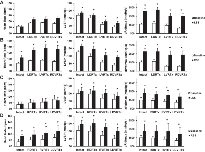
Fig. 6.
Hemodynamic data are presented as means ± SE. A: hemodynamic changes observed with left dorsal root transection-baseline compared with LSS. B: left dorsal root transection-baseline compared with RSS. C: right dorsal root transection-baseline compared with LSS. D: right dorsal root transection-baseline compared with RSS. *P < 0.05, significant difference from baseline with LSS and RSS.

