Fig. 7.

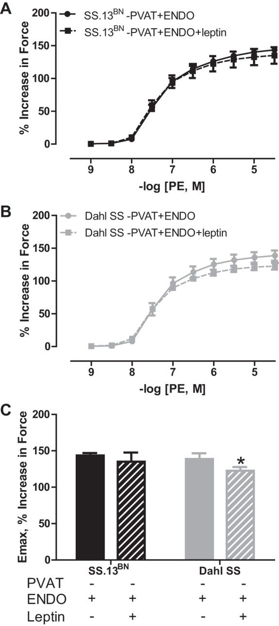
PE-induced vasoconstriction curves in aortic rings with the PVAT removed (−PVAT) and the endothelium intact (+ENDO) from SS.13BN rats (A) and Dahl SS rats (B) incubated in the presence or absence of leptin at 20 ng/ml for 20 min. C: maximum constriction (Emax) to −log [4.5, M] of PE. n = 5 rats/group. Number of multiple comparisons = 2. *P < 0.05 vs. Dahl SS aortic ring without leptin.
