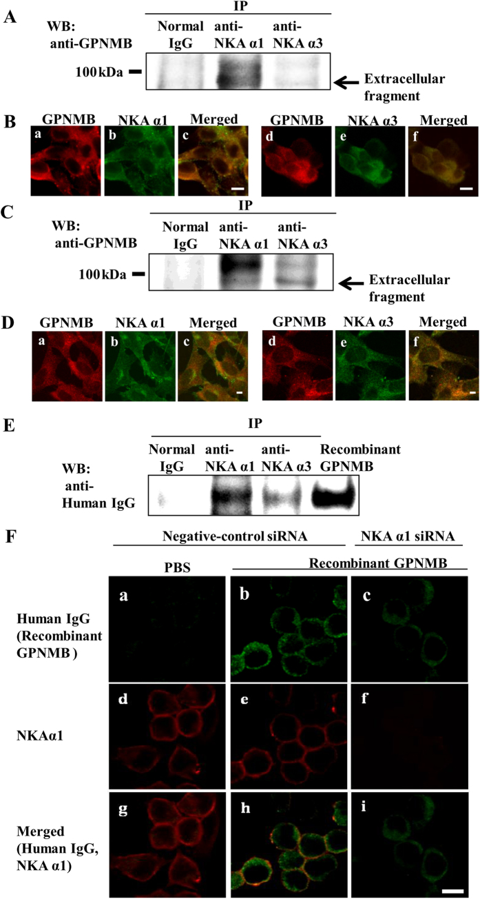
Figure 2. The extracellular fragment of GPNMB interacted and colocalized with the NKA α1 and α3 subunits in NSC-34 murine motor neuron cells.
(A) An equal amount of cell lysate was subjected immunoprecipitation (IP) by anti-NKA α1 antibody, anti-NKA α3 antibody, or normal mouse IgG (Normal IgG), and detected by western blot using anti-GPNMB antibody. (B) Co-immunostaining was conducted to localize NAK and GPNMB by anti-GPNMB antibody (red: a and d) and anti-NAK α1 antibody (green: b), or anti-NAK α3 antibody (green: e). (C) 661 W cells were plated at 2 × 10⁵ cells/well in 6-well plates and incubated overnight. The cell lysate was subjected immunoprecipitation (IP) by anti-NKA α1 antibody, anti-NKA α3 antibody, or normal mouse IgG (Normal IgG), and detected by western blot using anti-GPNMB antibody. (D) 661 W cells were seeded at 5 × 10⁴ cells/well in a Lab-Tek II Chamber slide (Thermo Fisher Scientific) and incubated overnight. Co-immunostaining in 661 W cell was conducted to localize NAK and GPNMB by anti-GPNMB antibody (red: a and d) and anti-NAK α1antibody (green: b), or anti-NAK α3 antibody (green: e). (E) NSC-34 cells were pretreated with human recombinant GPNMB and subjected to IP by anti-NAK α1 antibody, anti-NAK α3 antibody, or normal mouse IgG (Normal IgG), and detected by western blot with anti-human IgG antibody. (F) Co-immunostaining was conducted to localize of human recombinant GPNMB (green: a, b, and c) and NAK α1 (red: d, e, and f) in NSC-34 cells. The cells were pretreated with negative-control siRNA, and then treated with PBS (a, d, and g) or human recombinant GPNMB (b, e, and h). The cells were pretreated with NAK α1 siRNA, and then treated with the human recombinant GPNMB (c, f, and i). IP: immunoprecipitation, WB: Western blotting. Scale bar = 5 μm.

