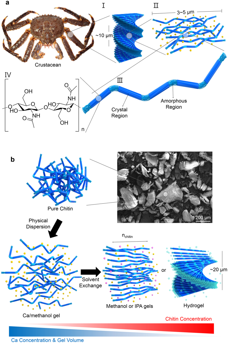
Figure 1. Hierarchically-ordered chitin microstructure in an arthropod cuticle.
(a) (I) Plywood structure of chitin nanofibrils, (II) chitin nanofibrils in the matrix (CaCO3 or proteins), (III) crystalline and amorphous domains of chitin nanofibril structure, and (IV) chitin structural formula. (b) Calcium-saturated methanol disintegrates chitin nanofibrils with minimal chemical modification, generating a Ca-methanol gel (disordered) (bottom-left panel). Ca2+ are removed from the Ca-methanol gel by washing with alcohol (methanol or IPA) and DI water, thus generating alcohol gels (methanol gel or IPA gel) in the N phase (bottom-middle panel) and a hydrogel in the N* phase (bottom-right panel). The yellow, pink, and blue beads represent three different types of solvent molecules: methanol-solvated Ca2+, alcohol (methanol or IPA), and water.

