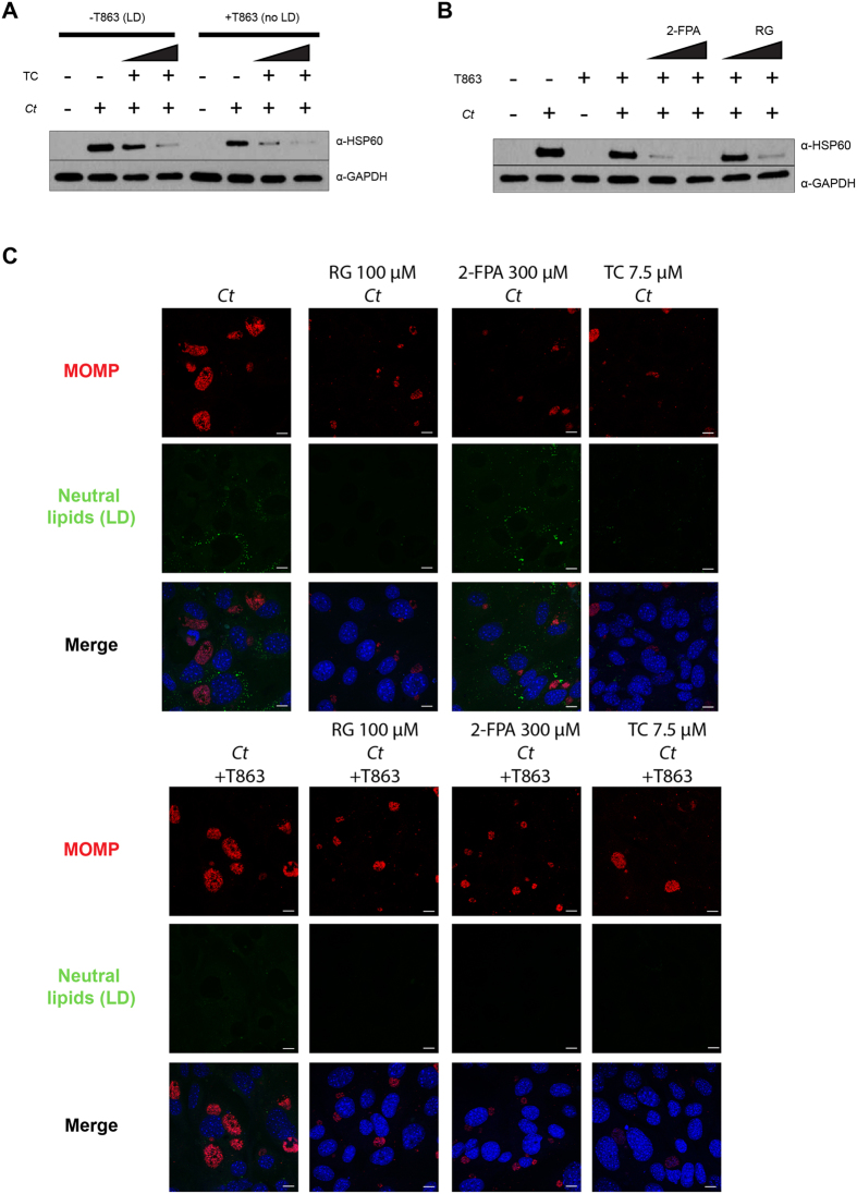
Figure 6. The block in C. trachomatis (Ct) inclusion growth through the inhibition of ACSLs is not dependent on lipid droplets (LD).
TC (A), 2-FPA and RG (B) were added to control and T863 treated DGAT2−/− MEF cells at two different concentrations (TC: 5 μM and 7.5 μM; 2-FPA: 200 μM and 300 μM; RG: 50 μM and 100 μM) for 16 h. The cells were then infected with Ct L2 without removing the inhibitor from the media. After 24 hpi, samples were prepared for WB analysis. Membranes were probed with anti-Ct HSP60 antibody and anti-human GAPDH antibody as a loading control. (C) DGAT2−/− MEF cells were treated with the inhibitor T863 (lower panels) or left untreated (upper panels). After 8 h of incubation, the following ACSL activity inhibitors were added: 100 μM RG, 300 μM 2-FPA, and 7.5 μM TC. After 16 h of incubation, cells were infected with Ct L2, without removing the inhibitors from the media. After 24 hpi, cells were fixed and visualized by confocal microscopy. Cells were stained with anti-Ct MOMP antibody (red), BODIPY 493/503 for neutral lipids (green), and Hoechst for nuclear and bacterial DNA (blue). Scale bar, 10 μm.

