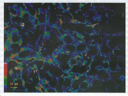
Figure 1.

Fluorescent reactions to lipopolysaccharides in cultured intrahepatic bile duct epithelial cells. Positive reactions were obvious in the cytoplasm. The color bar in the bottom on the left side shows the fluorescent intensities (the highest is red and the lowest green).
