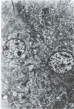
Figure 1.

Electron microscope observation of resected specimen 125I anti alpha fetoprotein treatment. (Li, male, 27 years old, HCC, admittance No.10206 tumor cross section, necrotic material, capsule intact electron microscope: Degeneration, necrosis of tumor tissue). Normal liver cell, from juxta tumor. × 2200
