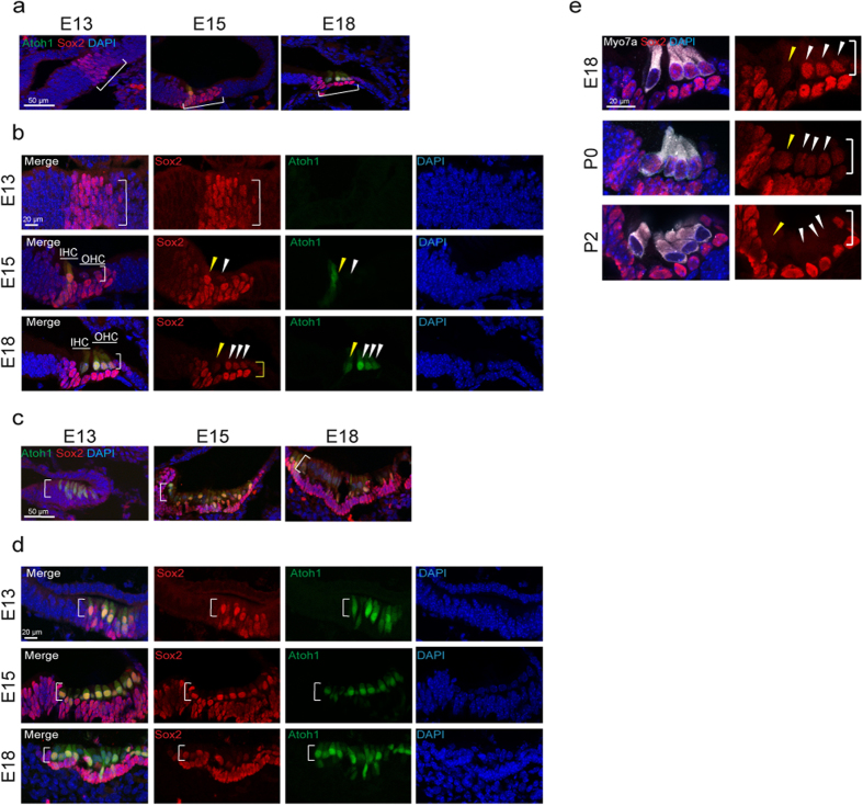
Figure 2. Sox2 Expression in the Developing Cochlea and Vestibular System.
(a) Sox2 (red) was expressed in the prosensory cells of the cochlea at E13 (bracket). Sox2 was expressed in the developing sensory epithelium at E15 and E18. (b) Atoh1-nGFP-positive cells (green) were first found in the sensory epithelium at E15 (bracket and arrowheads). Inner hair cells (IHC, yellow arrowhead), which develop before outer hair cells (OHC, white arrowheads), expressed Sox2 when newly formed at E15, and the newly formed inner and outer hair cells continued to express Sox2 at E18. Sox2 expression in hair cells had begun to decrease at that time (arrowheads) but was strong in supporting cells (yellow bracket). The mid-basal region is shown. (c) Atoh1-nGFP positive cells could be seen in the developing vestibular system as early as E13 (white bracket) and the cells expressed Sox2. (d) Atoh1-nGFP positive cells continued to express Sox2 at E15 (white bracket). Atoh1-nGFP-positive hair cells showed reduced Sox2 expression at E18 (white bracket). (e) Cochlear hair cells expressed myosin VIIa (Myo7a, white) at E18, P0 and P2. Sox2 was still expressed in hair cells in the cochlea until P0, but was undetectable in these cells at P2. It continued to be expressed in supporting cells at P2 (bracket and arrowheads).

