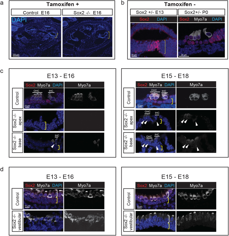
Figure 3. Sox2 is Required for Differentiation of Cochlear and Vestibular Hair Cells.
(a) The structure of the cochlea was intact when Sox2 was deleted by tamoxifen administration at E13 and analyzed at E16 (a Cre-negative Sox2flox/flox control was compared to a Sox2-Cre-ER; Sox2flox/+ knockout, Sox2 −/−). (b) The prosensory progenitors were present at E13, and hair cells had formed normally at P0 in an undeleted Sox2-Cre-ER; Sox2flox/+ mouse (Sox2 +/−). (c) After deletion of Sox2 at E13, the cochlea showed a nearly complete absence of hair cells at E16 (no hair cells were seen in the mid and apical regions and one row of dysmorphic inner hair cells was observed at the base). The position of myosin VIIa-positive (white) inner and outer hair cells are indicated by lines labeled IHC and OHC, and the position of the supporting cell layer is indicated by the yellow bracket (note the absence of Sox2 in the tamoxifen-treated animal). One inner and two outer hair cell rows at the base and a small number of dysmorphic hair cells in the apical region (arrowheads) were apparent at E18 when Sox2 was deleted 2 days later at E15. (d) After deletion of Sox2 at E13, the vestibular organs had weakly myosin VIIa-positive (white), dysmorphic hair cells at E16 in a layer (white bracket) above the supporting cell layer (yellow bracket; compare to the Sox2-positive (red) supporting cells and myosin VIIa/Sox2 double-positive hair cells in control). A greater number of hair cells were observed (white bracket) at E18 after deletion of Sox2 at E15. Nuclei were stained with DAPI (blue).

