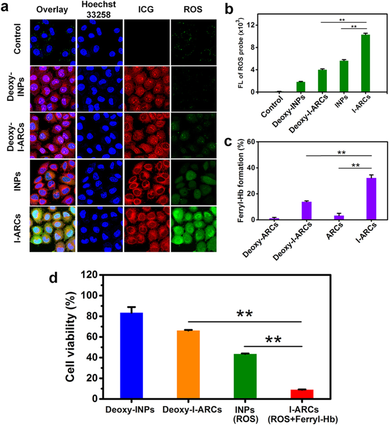
Figure 4. The boosted PDT effect of I-ARCs for cancer cell in vitro.
Cellular ROS detection after PDT measured by (a) confocal microscopy and (b) flow cytometry. Due to the oxygen-loaded Hb presence, I-NARCs generated abundant ROS that could produce oxidative damage in the cells. *P < 0.05, **P < 0.01. (c) Quantitative test of ferryl-Hb in the cell homogenate. *P < 0.05, **P < 0.01. (d) The cell viability was detected at 24 h cell incubation after laser treatments.

