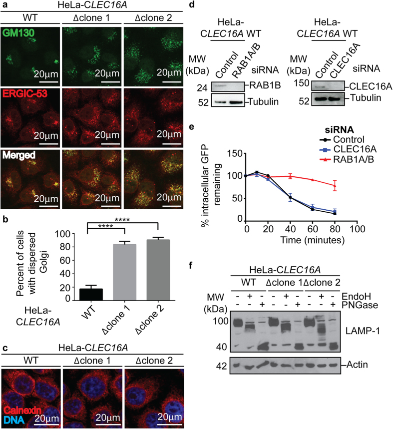
Figure 6. Structure and function of the Golgi apparatus in Clec16a-deficient cells.
(a–c) Confocal microscopy images of HeLa-CLEC16A cells stained for GM130, ERGIC-53 (a) and calnexin (c) and quantification of percent of cells/field with dispersed Golgi apparatus morphology (b) (representative of n = 3 experiments, minimum 60 cells/genotype quantified/experiment, data represent mean+/− s.e.m. and were analyzed by unpaired Student’s t-test; ****P < 0.0001). (d,e) A representative western blot for CLEC16A and Rab1b protein in HeLa C1 transfected cells utilized in hGH-GFP secretion assay (d). Remaining intracellular GFP (relative to time = 0) in HeLa C1 cells transfected with control, CLEC16A or RAB1A/B siRNA (E) (representative of n = 2 experiments). (f) A representative western blot of LAMP-1 in EndoH and PNGase digested HeLa-CLEC16A cells (representative of n = 4 experiments).

