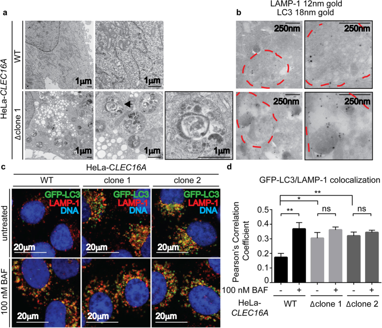
Figure 7. Clec16a-deficient HeLa cells have increased LAMP-1 and LC3 labeled single membrane bound structures.
(a) Transmission electron microscopy images of HeLa-CLEC16A cells (representative of n = 3 experiments). Black arrow indicates membrane bound structures containing cellular debris. (b) Cryo-immunoelectron microscopy images of immunogold labeling for LAMP-1 (12 nm gold) and LC3 (18 nm gold) in HeLa-CLEC16A cells (representative of n = 3 experiments). Red dotted lines indicate membrane bound structures containing cellular debris. (c,d) HeLa-CLEC16A cells, untreated or treated with 100 nm bafilomycin A1 (BAF), were imaged by confocal microscopy (c) and colocalization of GFP-LC3 and LAMP-1 was evaluated by Pearson’s Correlation Coefficient (d) (representative of n = 5 experiments, data represent mean+/− s.e.m. and were analyzed by unpaired two-tailed Student’s t-test; *P > 0.5, **P > 0.01; ns = not significant).

