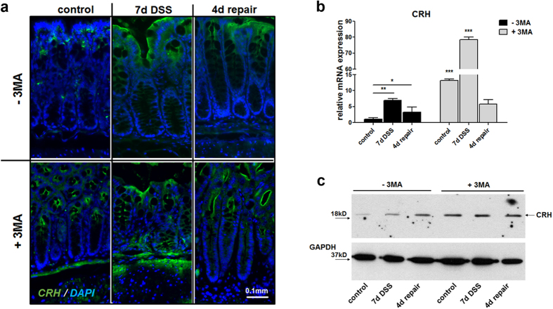
Figure 3. Autophagy inhibition positive regulates Crh expression and Crh protein levels.
(a) Representative images from immunohistochemistry in colon from wt mice under baseline conditions (control), following 7 days DSS administration and following 4d repair, indicating increased Crh in 3MA-treated mice. Sections were stained for Crh (green) and DAPI (blue). (b) Quantitative RT-PCR for mCRH conducted on colon homogenates from wt and Crh−/− under baseline conditions (control), during (7d DSS) or 4 days after (4d repair) 3% DSS administration. *p < 0.05, **p < 0.01, ***p < 0.001 (c) Western blot analysis of CRH protein levels. Whole protein lysates were extracted from wt and Crh −/− colons under baseline conditions (control), during (7d DSS) or 4 days after (4d repair) 3% DSS administration with or without 3MA administration.

