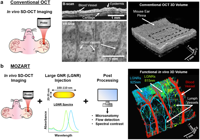
Figure 1. Overview of MOZART and its in vivo imaging capabilities.
(a) Conventional OCT scan of the mouse pinna shows micro-anatomic structures in 2D (B-scan), 2D en face slice, and volumetric rendering. The dashed lines on the volumetric rendering of the OCT structure show the locations of the B-scan and en face slice. (b) MOZART combines SD-OCT with large GNRs (LGNRs) as contrast agents that are detected with custom adaptive post-processing algorithms. This approach can be used to create images that contain additional functional information in vivo. The MOZART image reveals subcutaneously-injected LGNRs with two different spectra (green and cyan) draining into lymph vessels as well as flow in blood vessels (overlay in red). The conventional OCT and MOZART 3D images depict the same region (each volume is 4 mm × 4 mm × 1 mm).

