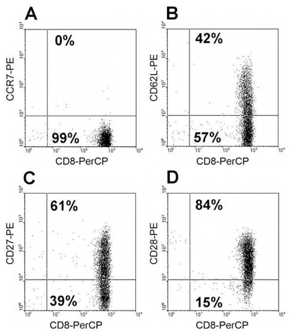
Figure 1.
Representative profile of memory phenotype marker expression on CD8+ T-cells in an IL-2-TIL sample from a melanoma patient. The IL-2-TIL sample was initiated from a melanoma patient in the presence of high-dose interleukin-2 (IL-2), and the expression of memory phenotype markers on T-cells was determined by flow cytometric analysis after staining with mAbs against human CCR7, CD27, CD28, CD62L and CD8. The x-axis represents the fluorescence staining with anti-CD8-PerCP, and the y-axis represents staining with anti-CCR7-PE (A), anti-CD62L-PE (B), anti-CD27-PE (C), and anti-CD28-PE (D), respectively.

