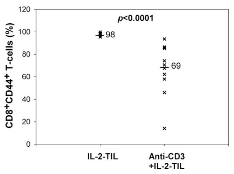
Figure 5.

Comparison of CD44 expression on T-cells between IL-2-TIL microcultures and anti-CD3+IL-2-TIL samples from melanoma patients. The IL-2-TIL microcultures refer to the TIL samples which were initiated from the tumor specimens resected from melanoma patients in the presence of high-dose interleukin-2 (IL-2) for two or three weeks. Anti-CD3+IL-2-TIL samples refer to the TIL samples after using the rapid expansion protocol in the presence of high-dose interleukin-2 (IL-2), anti-CD3 antibody and allogeneic peripheral blood monocytes for two weeks. T-Cells from both IL-2-TIL microculture samples and anti-CD3+IL-2-TIL samples were co-stained with R-phycoerythrin (PE)-conjugated anti-human CD44 and fluorescein isothiocyanate (FITC)-conjugated anti-human CD8 for flow cytometric analysis. The percentage of CD8+CD44+T-cells from both IL-2-TIL microculture samples and anti-CD3+IL-2-TIL samples were calculated and compared.
