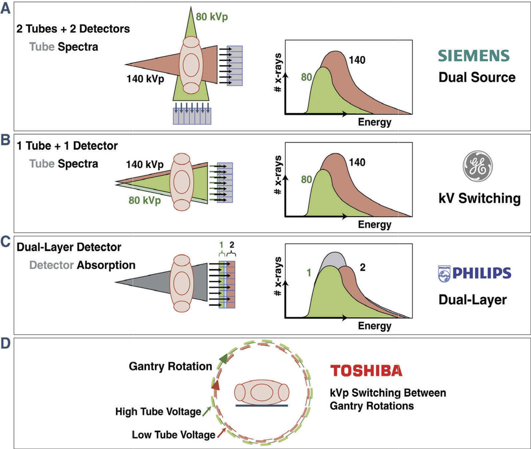
Figure 2. Schematic illustration of four different vendor-specific approaches for obtaining dual-energy Information.
(A) Dual source–detector pairs with each tube operating at a different voltage. Each x-ray source covers a different scan field. (B) Single source–detector pair with the source capable of rapid voltage switching (0.25 ms) in a single gantry rotation. (C) Single source–detector pair with a dual-layer detector made of two different materials capable of differentiating between low-energy (upper layer) and high-energy (bottom layer) photons, with the source operating at constant tube voltage allowing for dual-energy data during conventional CT scanning. (D) Single source–detector pair with tube voltage alternating between high and low kV with each gantry rotation. Reprinted with permission from the American College of Cardiology, Journal of the American College of Cardiology: Cardiovascular Imaging.[7] (Figures 2A, B, and C, Courtesy Philips Healthcare, Best, the Netherlands).

