Figure 10.
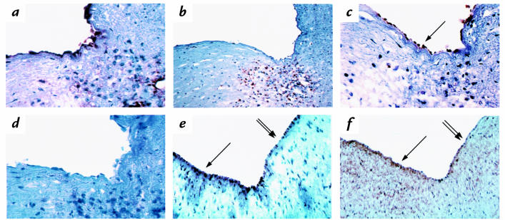
Human coronary lesions stained for CD 31, HAM 56, VCAM-1, 90.45, and HUTS-21. Human coronary lesions were stained with CD 31 (a), HAM 56 (b), 90.45 (c), or VCAM-1 (d). Antibodies in a–d were viewed with ABC and AEC. These four panels show that sections containing macrophages display endothelial CS-1 as detected by the 90.45 antibody but not VCAM-1 staining. In a separate study, the luminal endothelium of coronary vessels was stained for 90.45 (e) and HUTS-21 (f). Antibodies in e and f were viewed with DAB. Areas that stained most positively for 90.45 wer e mirrored by HUTS-21 (arrow), whereas areas of lesser staining were also parallel between the two antibodies (double arrow). ×1000. DAB, diaminobenzidine; ABC, avidin/biotinylated horseradish peroxide macromolecular complex; AEC, amino-9-ethyl carbazole.

