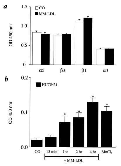
Figure 6.
MM-LDL increased the activation of β1 without increasing the total amounts of α5β1 on the apical surface. HAEC were either untreated or treated with MM-LDL for 4 h. ELISA was performed on nonpermeabilized cells to detect the apical expression of α5, α3, β3, and β1. MM-LDL did not increase the total levels of integrins on the surface of the HAEC (a). Values represent mean ± SD (n = 4). Using HUTS-21 to detect the activated form of the β1 integrin, ELISA demonstrated that MM-LDL increased the amount of β1 activation in a time-dependent fashion (b). Values represent mean ± SD (n = 4). *P < 0.0001. Each experiment is representative of four separate studies.

