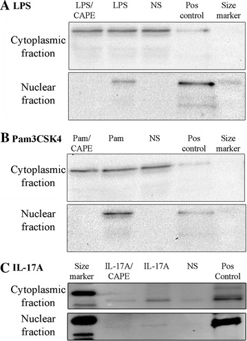
Figure 2.

Western analysis of NF-kB p65 in nuclear and cytoplasmic extracts of bTEC stimulated with LPS, Pam3CSK4 or IL-17A. NF-kB p65 was localized to the cytoplasm of non-stimulated cells (NS), but showed translocation to the nucleus following treatment of the cells with either LPS (panel A), Pam3CSK4 (Pam, panel B) or IL-17A (panel C). However, pre-treating the cells with the NF-κB inhibitor CAPE (10 μM) prior to exposure to agonists fully abrogated nuclear translocation of NF-kB p65 in the studies involving LPS and Pam3CSK4 (panels A and B), and partially in the studies involving IL-17A (panel C). Pos control: positive control, NF-kB p65. The findings were similar when the experiments were repeated using cells from different animals.
