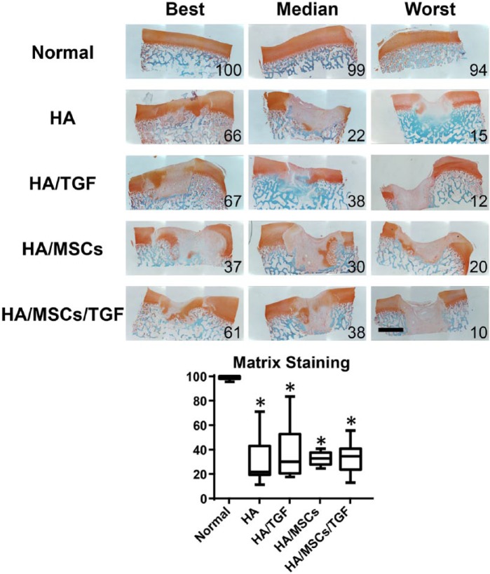
Figure 4.
Histological staining (Safranin O/fast green) for proteoglycans (red) and collagens (green) of full thickness cartilage defects treated with hyaluronic acid (HA) hydrogels, microspheres containing transforming growth factor–β (TGF), and mesenchymal stem cells (MSCs) showing entire defect and adjacent normal tissue. Numbers represent overall histological score for that specimen (*P < 0.05 vs. normal; scale bar = 2 mm).

