Figure 1.
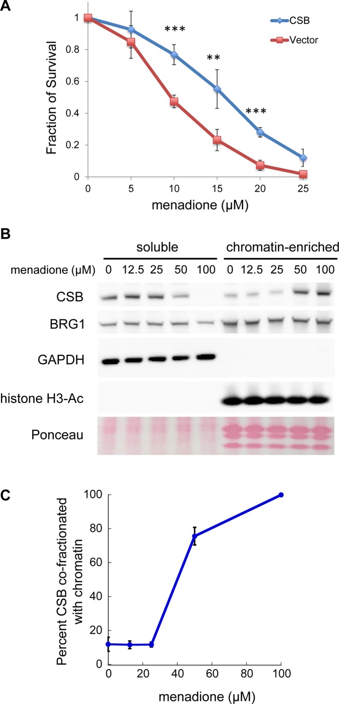
Menadione sensitivity assays. (A) CS1AN-sv cells were reconstituted with CSBWT or an empty vector. Stable cell lines expressing transgenes were assayed for viability 24 h after a 1-h menadione treatment with the indicated menadione concentrations. Shown are means ± standard errors of the mean (SEM) (n = 5). A paired t-test was used to determine if the difference in menadione sensitivity of CS1AN cells before and after CSB add-back was significant. Triple asterisks indicate P values < 0.001, and double asterisks indicate P values < 0.01. (B) Analysis of CSB partitioning in cells after a 1-h menadione treatment, with menadione concentrations as indicated. Western blots were probed with antibodies as noted. BRG1 was used as a loading control. GAPDH and acetylated histone H3 were used as markers for soluble and chromatin-enriched fractions, respectively. Total core histones were visualized by Ponceau S staining. (C) Quantification of CSB levels in the soluble versus chromatin-enriched fraction. Shown are means ± SEM (n = 4).

