Figure 4.
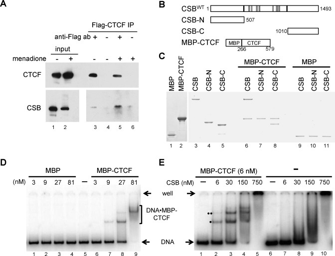
CSB interacts with CTCF in cells and in vitro. (A) Co-immunoprecipitation of CSB and CTCF in 293T transiently transfected with Flag-tagged CTCF, with or without a 1-h treatment of 100 μM menadione. 3.3% of the lysates used for IP were loaded as input. (B) Schematics of recombinant proteins used in (C–E). All CSB derivatives were N-terminally tagged with the Flag epitope. (C) Coomassie-stained gel showing that CSB directly interacts with CTCF. CSB-C, but not CSB-N, is sufficient for the CTCF association. MBP was used as a negative control. (D and E) EMSA assays showing that CSB enhances CTCF association with DNA. (D) Varying amounts of purified MBP-CTCF (lane 2 in C) or MBP (lane 1 in C) were incubated with a 32P-labeled, 200 bp DNA fragment containing a CTCF-binding motif (Supplementary Figure S4B). Protein–DNA complexes were resolved in a native 5% polyacrylamide gel. (E) Varying amounts of purified CSB were incubated with the radiolabeled DNA fragment in the presence or absence of MBP-CTCF. Reactions were subsequently resolved in a 5% native polyacrylamide gel. Protein–DNA complexes marked by ‘•’ and ‘••’ contain the MBP-CTCF protein, as they interacted with an anti-MBP antibody (Supplementary Figure S4A).

