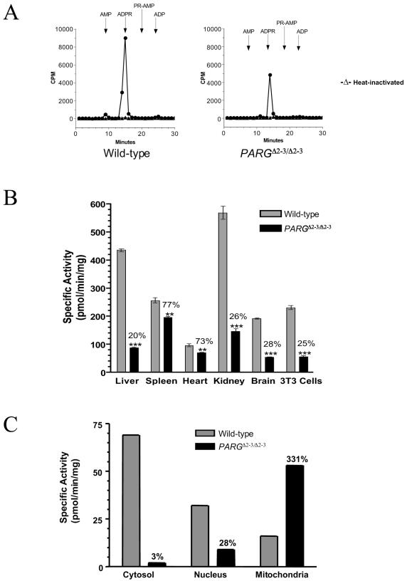
FIG. 3.
pADPR-degrading activity in PARGΔ2-3/Δ2-3 mutant mice and cells. (A) Liver samples extracted from wild-type and PARGΔ2-3/Δ2-3 mice were incubated with [32P]ADP-ribose polymers. Materials released from ADP-ribose polymers were analyzed by HPLC. The elution times for AMP, ADP-ribose (ADPR), phosphoribosyl-AMP (PR-AMP), and ADP are indicated. (B) PARG activity was measured in different tissues as well as in 3T3 EFs derived from mutant mice by a TLC assay. The results shown represent the mean and standard error of three determinations. The values demonstrate a significant difference between wild-type and PARGΔ2-3/Δ2-3 tissues and cells at P = 0.01 (***) or 0.05 (**). In all cases, the identity of the material released as ADP-ribose was verified by HPLC, as shown in panel A above. (C) Subcellular distribution of PARG activity was compared between wild-type and PARGΔ2-3/Δ2-3 EFs.

