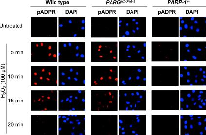
FIG. 4.
Synthesis and degradation of ADP-ribose polymers in PARG110-mutant cells. In situ immunodetection of poly(ADP-ribose) in wild-type and PARGΔ2-3/Δ2-3 cells by fluorescence microscopy is shown. Wild-type and PARGΔ2-3/Δ2-3 EF cells were treated or not with 100 μM H2O2 and stained with a polyclonal antibody for detection of ADP-ribose polymers. As a control, PARP-1−/− cells were treated in parallel. 4′,6′-Diamidino-2-phenylindole (DAPI) staining was used to visualize cell nuclei.

