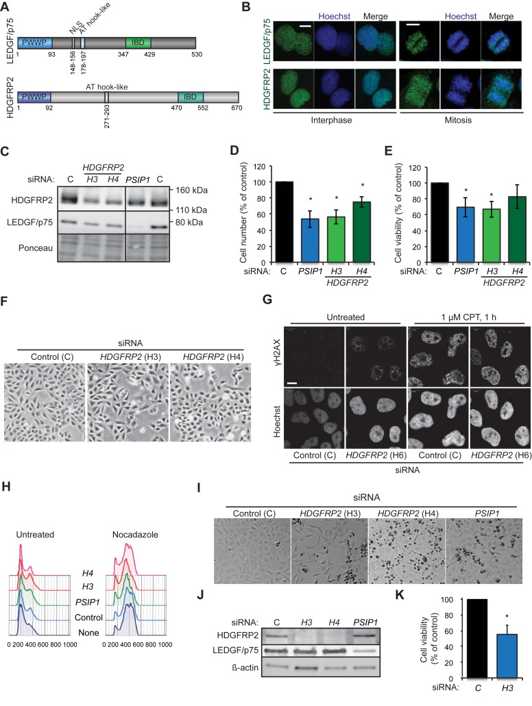
Figure 1.
HDGFRP2 is essential for the survival of U2OS and HeLa cells. (A) Schematic representation of the protein structures of LEDGF/p75 and HDGFRP2. (B) Representative confocal images of interphasic (left) and mitotic (right) U2OS cells fixed and stained for LEDGF/p75 and HDGFRP2. Scale bars, 10 μm. (C) Representative immunoblots of indicated proteins from lysates of U2OS cells transfected with non-targeting control (C), H3 and H4 (HDGFRP2) and PSIP1 (LEDGF) siRNAs for 72 h. Ponceau staining is shown as a control for equal loading. The vertical line indicates where the blot was cut to remove non-essential bands after staining and exposure. (D and E) The number (D) and viability (E) of U2OS cells transfected as in (C) were analysed by Celigo cytometer and PrestoBlue assay, respectively. (F) Representative brightfield images of U2OS cells transfected as in (C). (G) Representative confocal images of U2OS cells transfected with non-targeting control (C) or H6 (HDGFR2) siRNAs for 72 h and left untreated or treated with 1 μM camptothecin (CPT) for 1 h. DNA damage foci were detected by staining for γH2AX and nuclei were stained with Hoechst. Scale bars, 10 μm. See Supplementary Figure S1 for an immunoblot demonstrating the efficacy of H6 siRNA. (H) Cell cycle profiles of U2OS cells transfected as in (C) and left untreated (left) or treated with 4 μg/ml nocodazole for the last 24 h (right) before fixation and staining with propidium iodide (PI). PI staining was quantified using flow cytometry and analysed by FlowJo software. (I and J) Representative brightfield images of HeLa cells transfected with indicated siRNAs for 72 h (I) and immunoblots of proteins extracted from the same cells (J). Note an increase in condensed dead cells (small and dark) in samples treated with HDGFRP2 and PSIP1 siRNAs. (K) The viability of HeLa cells transfected with indicated siRNAs for 72 h was analysed by PrestoBlue assay. Error bars, SEM of two (D and E) or three (K) independent triplicate experiments. *P < 0.05 when compared with cells transfected with control siRNA in parallel.

