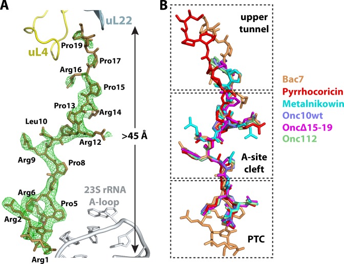
Figure 2.
The structure of PrAMPs in their ribosome-bound conformation. (A) Structure of Bac71–35 bound inside the peptide exit tunnel of the ribosome. The difference Fourier map calculated at 3.0 Å resolution using initially phased diffraction data (Fobs–Fcalc, green) contoured at ∼3σ shows clear unbiased electron density for 19 residues of Bac71–35. The 23S rRNA A-loop and ribosomal proteins uL4 and uL22 are shown. (B) The superposition of all PrAMPs bound to the ribosome reveals that the common core (middle) region overlaps almost perfectly. Bac71–35 is colored brown, Onc112 is green [PDB ID:4Z8C (27)], Metalnikowin is cyan, Onc10wt is slate, OncΔ15–19 is magenta and Pyrrhocoricin is red.

