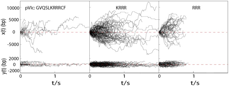
Figure 1.
Diffusion of TMR-pVIc, TMR-KRRR and TMR-RRR along flow stretched λ-DNA molecules in 2 mM NaCl buffer. x(t) and y(t) are the displacements along and transverse to λ-DNA, respectively. For N-terminal labeled peptides, no significant change in D1 was observed between pH 6.5 and pH 7.4, and thus, diffusion trajectories observed at pH 6.5 and pH 7.4 were combined to give more accurate estimation of D1. Dotted horizontal lines indicate missing points resulting from dye blinking.

