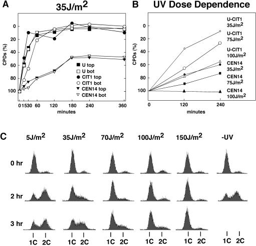
FIG. 7.
UV dose dependence of centromere repair and cell growth after UV damage. (A) Inhibition of CEN14 repair after irradiation with 35 J/m2. The fraction of CPDs in CEN14 and flanking regions is shown (U and CIT1) after exposure to a low dose of UV (35 J/m2) and incubation at room temperature in the dark for up to 360 min. Chromosomal regions U and CIT1 around CEN14 are shown (as in Fig. 1). bot, bottom strand; top, top strand. (B) Fraction of CPDs in CEN14 after irradiation at 35, 75, and 100 J/m2 and incubation in the dark at room temperature for 120 and 240 min. Data are for the bottom strand. (C) Cells arrested with α-factor in G1 were released by washing in SD without amino acids, irradiated at 5 to 150 J/m2 or left unirradiated (−UV), and after addition of the appropriate amino acids incubated in the dark at room temperature for up to 3 h. Cells were fixed, stained with propidium iodide, and analyzed by flow cytometry as described previously (35). 1C and 2C, DNA content of haploid cells before and after replication, respectively.

