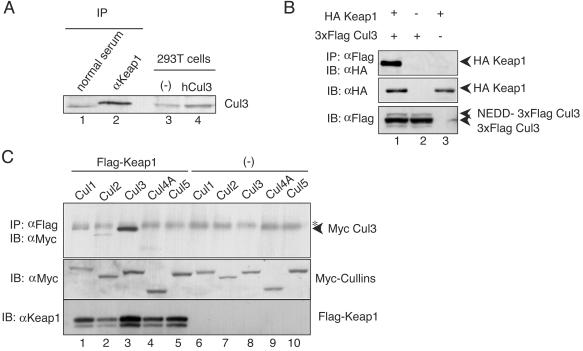
FIG. 4.
Keap1 associates specifically with Cul3 in vivo and in vitro. (A) Complex formation of Keap1 and Cul3 in 293T cells. Endogenous Keap1 was precipitated with anti-Keap1 antibody and protein G beads (IP). The immunocomplex was subjected to immunoblot analysis with anti-Cul3 antibody. Whole-cell extracts of 293T cells expressing human Cul3 were used as a control (lanes 3 and 4). (B) Association between Keap1 and Cul3 in a transient-expression system. Whole-cell extracts prepared from 293T cells transfected with expression plasmids of HA-tagged Keap1 (1 μg) and 3xFlag Cul3 (1 μg) were subjected to immunoprecipitation (IP) with anti-Flag (M2) beads and immunoblot analysis with anti-HA antibody (IB). Analyses of cells expressing 3xFlag Cul3 with or without HA-Keap1 (lanes 1 and 2) are shown. Lane 3 is loaded with cell extracts expressing HA-Keap1 alone. (C) Among the Cul family proteins, Cul3 specifically interacts with Keap1. Expression plasmids (1 μg each) of Cul1 (lanes 1 and 6), Cul2 (lanes 2 and 7), Cul3 (lanes 3 and 8), Cul4A (lanes 4 and 9), and Cul5 (lanes 5 and 10) were transfected into 293T cells in the presence (lanes 1 to 5) or absence (lanes 6 to 10) of Flag-fused Keap1. Immunoprecipitation and immunoblot analyses were performed as described above (top). The asterisk indicates a nonspecific band. The expression levels of Cul proteins and Flag-Keap1 were verified by immunoblot analysis with anti-Myc and anti-Keap1 antibodies (middle and bottom, respectively)

