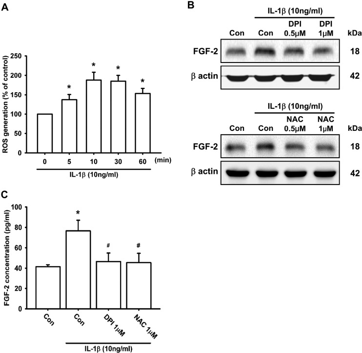
Figure 3. ROS generation is involved in IL-1β-induced FGF-2 expression.
ATDC5 cells were labelled with 10 μM H2DCFDA, and then incubated with IL-1β for the indicated times. The fluorescent intensity was measured by flow cytometry (A; n=9). ATDC5 cells were pre-treated with DPI or NAC for 30 min, and then stimulated with IL-1β for 24 h. FGF-2 expression was examined by Western blotting (B; n=8) and ELISA (C; n=6). Quantification results are expressed as means±S.D. *P<0.05 compared with the 0 min group in (A) and compared with the Con group (control) in (C); #P<0.05 compared with the IL-1β-treated group in (C). Molecular masses are indicated in kDa. β-Actin was used as a loading control.

