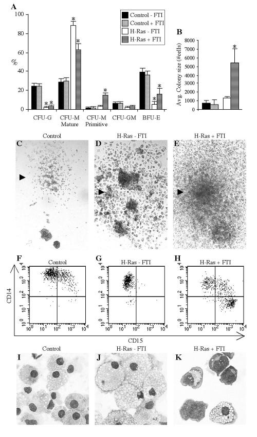
FIG. 3.
The amplitude of Ras signaling influences lineage selection and the proliferative capacity of hematopoietic progenitors. (A) Transduced cells were plated in CFC assays in the presence or absence of 50 nM FTI, as indicated. The mean percentages ± standard errors of CFU-G, primitive and mature CFU-M, and granulomonocytic and BFU-E progenitors scored on day 11 of culture (nine experiments) are shown. An asterisk indicates significant difference (P < 0.01) between results with H-Ras-expressing samples and their control counterparts. (B) The average size of colonies (±SD) generated by transduced progenitors in the presence or absence of FTI was determined by collecting all cells from day 12 methylcellulose plates, counting them, and dividing the cells by the number of colonies present. The results of three experiments, with two methylcellulose plates per condition per experiment, are presented. An asterisk indicates significant difference (P < 0.03) between results with H-Ras plus FTI and the control plus FTI. (C to E) Representative monocytic colonies on control, H-Ras without FTI, and H-Ras plus FTI plates (magnification, ×25). (F to H) Flow cytometric assessment of CD15 and CD14 expression on cells from representative single monocytic colonies on control, H-Ras without FTI, and H-Ras plus FTI plates. (I to K) MGG-stained cytospins of cells from representative monocytic colonies on control, H-Ras without FTI, and H-Ras plus FTI plates (magnification, ×400).

