Figure 2.

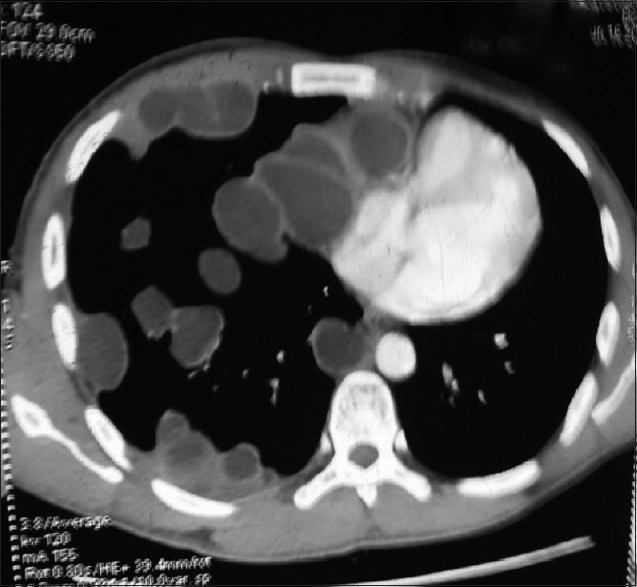
Multiple hydatidosis lungs. Computed tomography thorax (mediastinal window) showing multiple fluid-filled cystic lesion along the mediastinal and costal pleura and lung parenchyma

Multiple hydatidosis lungs. Computed tomography thorax (mediastinal window) showing multiple fluid-filled cystic lesion along the mediastinal and costal pleura and lung parenchyma