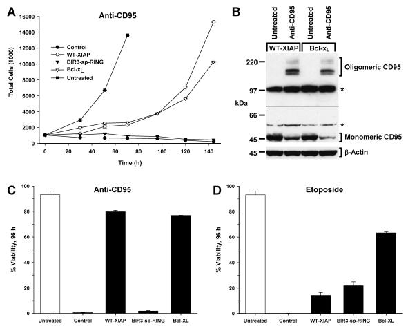
FIG. 2.
Protective properties of XIAP following long-term anti-CD95 treatment. (A) Jurkat-derived cells were treated with anti-CD95 for 6 days. Proliferation was assessed by cell density determination every 24 h using a Coulter Counter. Representative data from untreated control cells are shown (filled squares). (B) Lysates were prepared from WT-XIAP- and Bcl-xL-expressing Jurkat cells prior to and following treatment with anti-CD95 for 6 days and then immunoblotted for the presence of CD95. Shown are two separate exposures of the same immunoblot, separated by the black line: the lower exposure (1 min) shows monomeric CD95, and the upper exposure (5 min) shows oligomeric CD95. Note that while levels of monomeric CD95 decreased following treatment, the presence of oligomeric CD95 following treatment accounted for this loss. As a loading control, immunoblot analysis for β-actin was also performed (lower panel). (C and D) Viability of cell lines shown in panel A (C) or of cells treated with etoposide (D) was assessed 96 h following treatment by PI staining followed by flow cytometry. The average ± standard deviation of multiple independent measurements is shown. Data are representative of at least three independent experiments.

