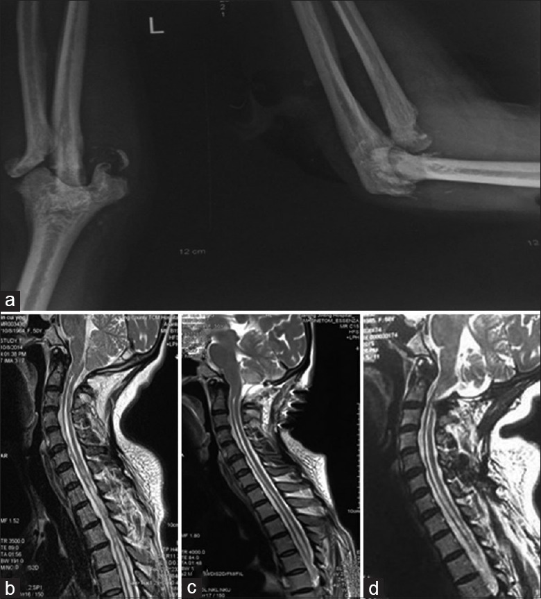
Figure 1.

X-ray radiograph of the left Charcot elbow joint and sagittal T2-weighted magnetic resonance imaging demonstrating the evolution of Chiari malformation associated with syringomyelia. (a) X-ray radiograph of the left elbow demonstrating destructive arthropathy and slight calcification. (b) Sagittal T2-weighted of preoperative magnetic resonance imaging demonstrating type I Chiari malformation with syringomyelia from cervical region to thoracic region. (c) Seven days after surgery demonstrating good cerebellar retraction and significant reduced syringomyelia. (d) Half a year after surgery demonstrating no significantly changed syringomyelia.
