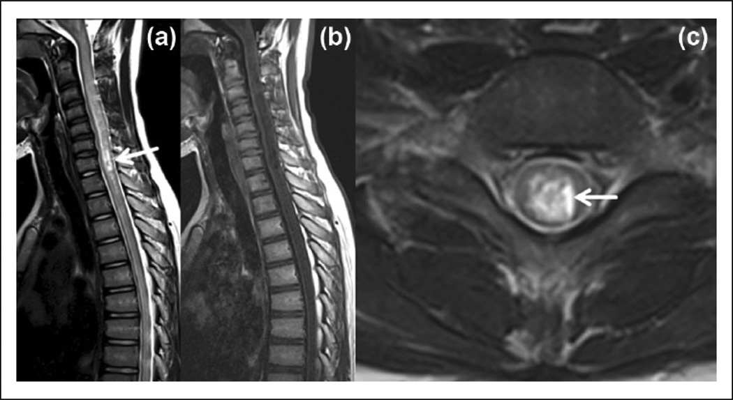
Figure 1.
Spinal MRI of a 12-year-old female with seropositive NMO shows: (a) A typical longitudinally extensive transverse myelitis extending into the brainstem with ‘bright spotty lesions’ (arrow) on a T2-weighted sagittal image. (b) The lesion is a very hypointense, ‘T1 dark’ on a T1-weighted image. (c) On a T2-weighted axial image, the lesion is both centrally- and peripherally-located and peripheral T2 hypointensity is preserved. Arrow demonstrates the ‘bright spotty lesion’.
MRI: magnetic resonance imaging; NMO: neuromyelitis optica.

