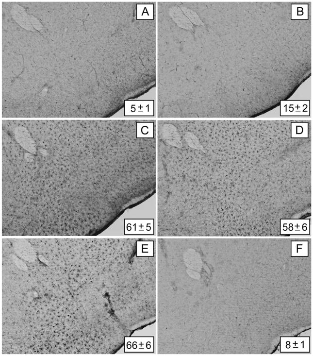
Figure 3.
Effects of clorgyline, L-DOPA, and reserpine on microglial activation in the nucleus accumbens (NAc) caused by a neurotoxic methamphetamine (Meth) regimen. Mice (n = 3–5 per group) were treated as described in the text and analyzed for microglial activation in the NAc 48 h after the last Meth injection. Microglia counts are presented as means ± SEM. Treatment conditions and microglia counts for each panel are (A) control (5 ± 1), (B) Meth (15 ± 2), (C) clorgyline + Meth (61 ± 5), (D) L-DOPA + Meth (58 ± 6), (E) reserpine + Meth (66 ± 6), and (F) reserpine only (8 ± 1). Significant differences were determined via one-way analysis of variance (ANOVA) followed by Tukey’s multiple comparison test: p < 0.001 for clorgyline + Meth, L-DOPA + Meth, and reserpine + Meth relative to control. No significant differences were determined for Meth or reserpine only relative to control (p > 0.05). Reprinted with permission from Thomas et al. (2009). The color image is available in the online posting of this article at www.ilarjournal.com.

