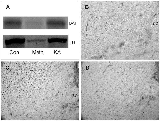
Figure 4.
Effects of kainic acid (KA) on dopamine (DA) nerve terminals and microglial activation. Mice (n = 3–5 per group) were injected intraperitoneally with kainic acid (25 mg/kg) or with a neurotoxic regimen of methamphetamine (Meth) (4 × 5 mg/kg, 2 hr between injections) and analyzed 48 hr after treatment for expression levels of the dopamine transporter (DAT) and tyrosine hydroxylase (TH) and for microglial activation. Levels of DAT and TH were determined in combined caudate-putamen/nucleus accumbens (CPu/NAc) samples by western blot analysis (A). Microglial activation was determined by histochemical staining using Isolectin B4. The anterior commissure (ac) is labeled for orientation. Very little microglial activation is seen in the NAc or CPu of control mice (B) whereas Meth treatment causes extensive microglial activation in the CPu but not in the NAc (C). Kainic acid does not lead to microglial activation in either the NAc or CPu (D). Con, control. The color image is available in the online posting of this article at www.ilarjournal.com.

