Figure 2.
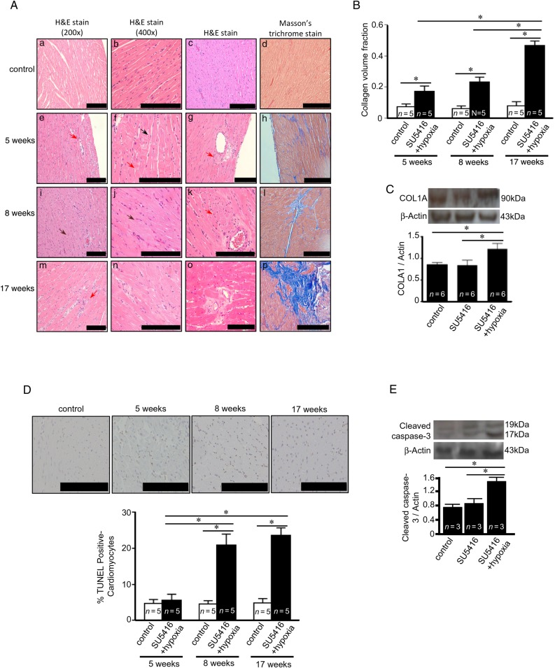
PAH causes apoptosis and fibrosis in the RV. Rats were subjected to SU5416/hypoxia followed by 2, 5, or 14 weeks of normoxia (5-, 8-, and 17-time points, respectively). (A) H&E stain images from two different magnifications. H&E staining of the perivascular region is also shown. Masson's trichrome stain with collagen stained in blue with 200× magnification. (B) Quantification of Masson's trichrome stain detecting collagen volume fraction. (C) Western blotting showing increased collagen type 1a (COLA1) in the RV at 8 weeks after the SU5416 injection. (D) TUNEL assays were performed and percentage of apoptotic cardiomyocytes was determined. (E) Western blotting showing increased cleaved caspase-3 at 8 weeks after the SU5416 injection. The symbol (*) denotes that the values are significantly different from each other at P< 0.05 as determined by one-way ANOVA. Results presented in (B) and (D) were also analysed using two-way ANOVA. Scale bars, 200 μm.

