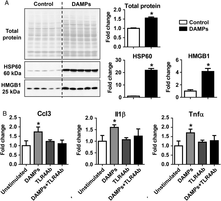
Figure 5.
DAMPs activated N1 neutrophil polarization in vitro via TLR4. (A) LV tissues that underwent freeze–thaw cycles (the DAMPs group) contained significantly higher total protein, HSP60, and HMGB1, compared with controls, as evaluated by immunoblotting. n = 5 LVs per group. The same volume of all samples was loaded (2 µL). (B) DAMPs stimulated the production of proinflammatory markers Ccl3, Il1β, and Tnfα, which was abolished by the anti-TLR4 neutralizing antibody. The data were expressed as fold change (normalized to control or unstimulated group). n = 3–6 mice per sample and n = 4–9 samples per group; *P < 0.05 vs. controls. One-way ANOVA was used.

