Figure 5.
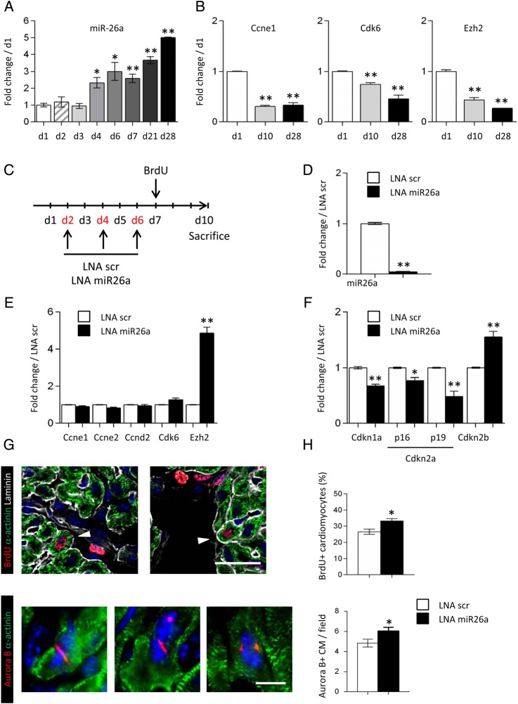
miR-26a regulates cardiomyocyte proliferation in the neonatal heart. (A) Time course of miR-26a expression in the postnatal mouse heart. (B) Expression of the miR-26a target genes, Ccne1, Cdk6, and Ezh2, in the mouse heart at d1, d10, and d28 of age. (C) Schematic representation of the protocol used to inhibit miR-26a in the neonatal mouse heart. (D) miR-26a expression in LNA scr (white bars) and LNA miR26a (black bars) treated hearts at 10 days of age. (E) Expression of miR-26a target genes in LNA scr (white bars) and LNA miR26a (black bars) treated hearts. (F) Expression of Ezh2 target genes, Cdkn1a, Cdkn2a, and Cdkn2b, in LNA scr (white bars) and LNA miR26a (black bars) treated hearts. Data are expressed as mean ± SEM; n ≥ 4; *P < 0.05, **P < 0.01. (G) Immunostaining of heart sections using anti-BrdU (red) anti-α-actinin (green) and anti-laminin (gray) antibodies (upper panel), or using anti-Aurora B (red) and anti-α-actinin (green) antibodies (lower panel). Nuclei were stained with DAPI (blue). Scale bar: 20 μm in upper panels and 10 μm in lower panels. (H) Quantification of BrdU-positive cardiomyocytes, and Aurora B-positive cardiomyocytes per ×40 magnification field in hearts of LNA scr (white bars) and LNA miR26a-treated (black bars) neonatal mice. Data are expressed as mean ± SEM; number of analysed cardiac sections for each group ≥30; **P < 0.01.

