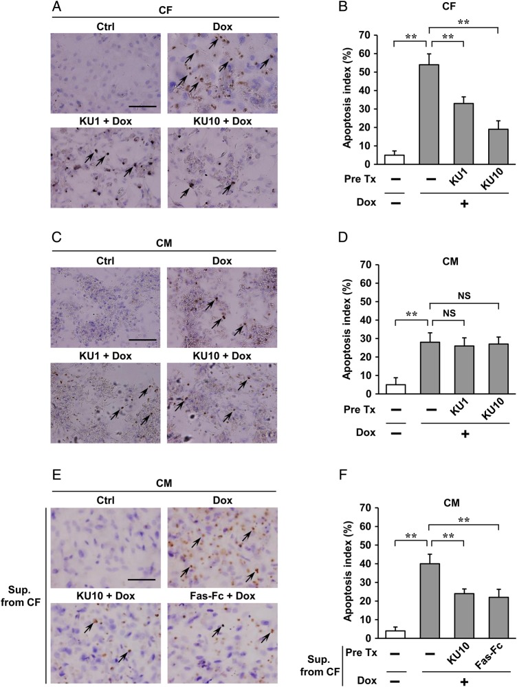
Figure 4.
Regulation of Dox-induced apoptosis by ATM in cardiac fibroblasts in vitro. (A–D) Effects of ATM inhibitor (KU55933) on Dox-induced apoptosis of cardiac fibroblasts (CFs) or cardiomyocytes (CMs) were assessed by TUNEL staining. Cardiac fibroblasts or cardiomyocytes isolated from neonatal rats were pre-treated with 1 or 10 µM KU55933 for 30 min followed by administration of 1 µM Dox for 24 h and then immunostained for TUNEL assay (brown). Haematoxylin was used as nuclear stain (blue). Arrows indicate representative TUNEL-positive cardiac fibroblasts (A) or cardiomyocytes (C). Scale bar, 100 µm. Apoptosis index (percentage of TUNEL-positive nuclei) was calculated as TUNEL-positive nuclei/total nuclei × 100 (%). Apoptosis index of cardiac fibroblasts (B) or cardiomyocytes (D) was plotted. (E and F) Effects of conditioned medium from cardiac fibroblasts on Dox-induced cardiomyocyte apoptosis were assessed by TUNEL staining. Cardiomyocytes isolated from neonatal rats were incubated for another 24 h in conditioned medium prepared from cardiac fibroblasts pre-treated with KU55933 (an inhibitor of ATM, 10 µM) or Fas-Fc (neutralizing agent of FasL, 10 µg/mL) 30 min followed by administration of 1 µM Dox for 24 h, and then immunostained for TUNEL assay (brown) in cardiomyocytes. Arrows indicate representative TUNEL-positive cardiomyocytes (E). Scale bar, 100 µm. Apoptosis index (percentage of TUNEL-positive nuclei) was calculated as TUNEL-positive nuclei/total nuclei × 100 (%). Apoptosis index of cardiac cells was plotted (F). The results are expressed as means ± SD (n = 3 per group, total of nine visual fields); **P < 0.01; NS: not significant, by one-way ANOVA followed by a post hoc Tukey's multiple comparisons test. Pre Tx, pre-treatment; KU1, KU55933 1 µM; KU10, KU55933 10 µM; Sup. From CF, conditioned medium prepared from cardiac fibroblasts; Ctrl, Pre Tx-free and Dox-free.

