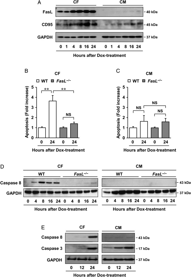
Figure 5.
Mechanism of FasL in regulating Dox cardiotoxicity. (A) Dox-induced FasL and CD95 expression as assessed by western blot analysis. Cardiac fibroblasts and cardiomyocytes isolated from neonatal mice were incubated in 1 µM Dox for indicated times. Cells were lysed and subjected to western blot analysis with indicated antibodies (n = 3 per group). (B and C) Effects of FasL on Dox cardiotoxicity in vivo. Cardiac fibroblasts (B) and cardiomyocytes (C) isolated from adult wild-type or FasL−/− mice were treated with 1 µM Dox for 24 h and then assessed using the Cell Death Detection ELISAPlus kit. The results are expressed as means ± SD (n = 3 per group); **P < 0.01; NS: not significant, by two-way ANOVA followed by a post hoc Tukey's multiple comparisons test. (D) Effects of FasL on the apoptosis-related factor caspase-8 in vivo. Cardiac fibroblasts and cardiomyocytes isolated from adult wild-type or FasL−/− mice were treated with 1 µM Dox for indicated times and then assessed by western blot analysis (n = 3 per group). (E) Dox-induced caspase-8 and -3 expressions as assessed by western blot analysis. Cardiac fibroblasts and cardiomyocytes isolated from neonatal mice were incubated in 1 µM Dox for indicated times. Cells were lysed and subjected to western blot analysis with indicated antibodies (n = 3 per group). GAPDH was used as the loading control.

