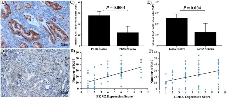
Fig 6. Immunohistochemical staining and correlation between PKM2, LDHA and Ki-67.
(A) Tumours with strongly positive PKM2 expression had most of the nucleus stained for Ki-67 (x100 magnification). (B) Tumours weakly positive for PKM2 had scant Ki67 staining (x100 magnification). (C) & (D) Correlation between PKM2 staining and Ki-67 positive cells. (E) & (F) Correlation between LDHA staining and Ki-67 positive cells. There was a significant correlation for both PKM2 and LDHA.

