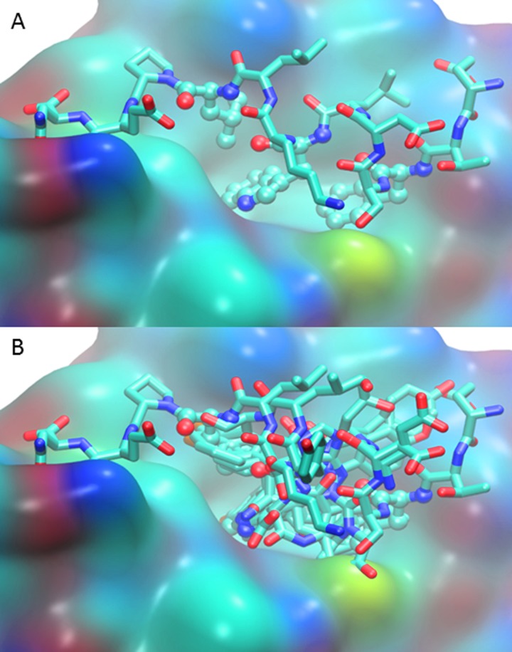
Fig 1.
(A) MDM2 binding interface (surface view with CPK atom coloring) with native p53 N-terminal peptide (licorice, also CPK coloring) bound in 1YCR crystal structure [15]. The three key binding residues, Phe19, Trp23, and Leu26, are highlighted with ball and stick view. (B) MDM2-bound p53 N-terminal peptide aligned with representative protein-bound inhibitors. For clarity the protein surface of only 1YCR is shown. The PDB ID and inhibitors included are 1YCR native p53 peptide [15], 1T4E benzodiazepinedione [33], 3LBL MI-63-analog [34], 3LBK imidazol-indole [34], 3JZK chromenotriazolopyrimidine [35], 4HG7 nutlin-3a [36], 4JRG pyrrolidine carboxamide [37], 4UMN stapled peptide [38].

