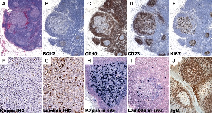
Fig 2. Immunohistochemistry of isolated atypical follicles.
An axillary lymph node dissection in a 58 year-old woman with breast carcinoma (case 3) shows a lymph node with a cluster of follicles (A). BCL2 expression is absent in both the involved and uninvolved follicles (B) and the involved follicle shows diminished CD10 expression relative to the surrounding normal germinal centers (C). CD23 demonstrates an intact follicular dendritic network around the involved follicle (D). Ki-67 is polarized in surrounding reactive follicles, but is not polarized in the involved follicles (E). The involved follicle in case 1 shows lambda light chain-restricted B-cells (F and G). Case 2 shows highly atypical large cells that by situ hybridization (ISH) for immunoglobulin kappa and lambda light chains show kappa-specific RNA in the majority of the atypical cells, confirming light chain restriction in the involved follicle (H and I). A periaortic lymph node from a 53-year old woman (case 4) shows abnormal strong IgM protein expression in the centroblasts of an involved follicle whereas the uninvolved follicle shows a weak dendritic pattern of IgM reactivity, which is typically seen in normal follicles (J).

