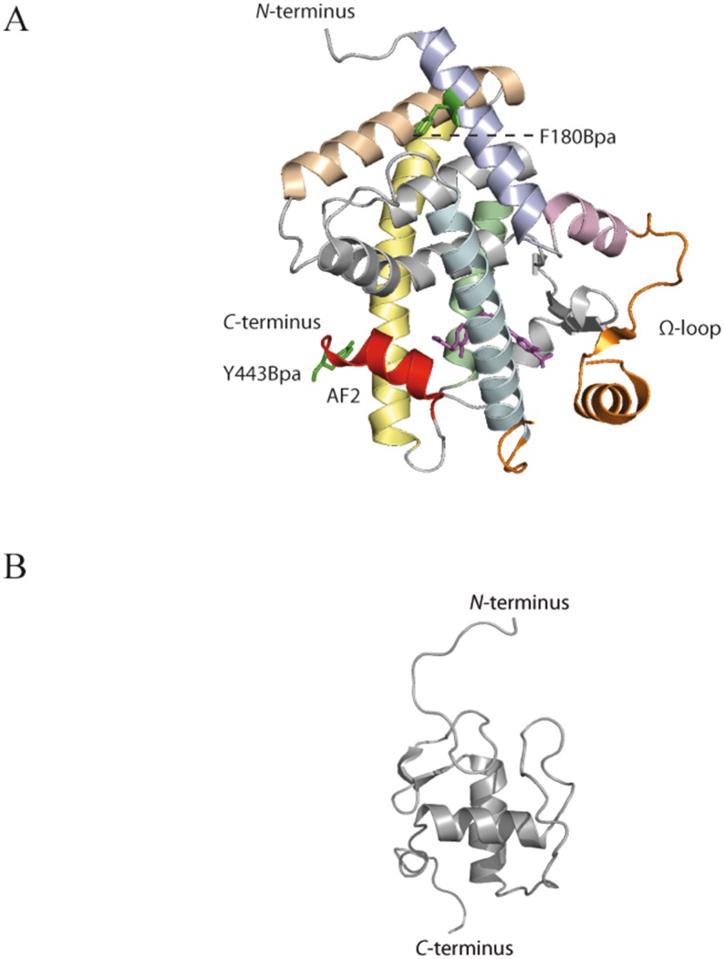
Fig 4. High-resolution 3D-structures of PPAR-β/δ.
(A) X-ray structure of PPAR-β/δ LBD (pdb 3TKM), bound to agonist GW0742; The activation function helix 2 (AF2, helix 12) is shown in red, the flexible Ω-loop is shown in orange; the amino acids replaced by Bpa are shown as green sticks; the ligand GW0742 is shown in stick representation in magenta. Helices containing amino acids that are involved in cross-linking are colored (helix 1: light blue; helix 2: light pink; helix 4: pale cyan; helix 8: pale green; helix 10: wheat; helix 11: pale yellow). (B) NMR structure of PPAR-β/δ DBD (pdb 2ENV).

