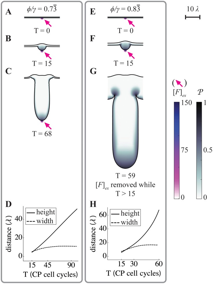
Fig 7. Formation of growing, constant-width fingers.
Spatial simulations similar to those in Figs 3–5 were carried out, using a larger domain size, slower diffusivity of feedback factors F and G, and incorporating strong uptake of G on the stromal side of the epithelium (for parameters see S1 Table; the characteristic decay lengths of F and G, denoted λ, were identical, and can be appreciated from the 10λ scale bar in the upper right corner). Boundary conditions were also taken to be periodic. Simulations all began from the same initial flat geometry, with an exogenous source of F in the center of the domain (Fex, arrows), but using different values of the endogenous feedback ratio (ϕ/γ), as shown. A-C. At the lower ϕ/γ ratio, a fingerlike structure elongates as long as the exogenous signal is present. Values of P are high primarily near the source of F. D. The length of the finger in A-C increases linearly in time, but after initial growth, its width remains constant. E-G. At the higher ϕ/γ ratio, a similar-looking structure is generated, but it continues to grow even after the exogenous source of F is removed after the 15th CP cell cycle. Values of P remain high primarily near the tip of the growing finger. H. Although the finger elongates at an approximately exponential rate, its width also stabilizes at a constant value. Distances in D and H are plotted in units of the intrinsic decay length of both F and G within the tissue (which is the square root of the ratio of diffusivity to the rate constant of uptake).

